Horse Lameness: Causes, Diagnosis, Treatment and When to Call the Vet
By FarmVetGuide Editorial Team · Published March 2026 · Updated March 2026 · Based on verified data from our directory of 9,500+ practices
A lame horse is a horse that cannot work, and in many cases, cannot be comfortable. Horse lameness is the most common cause of reduced performance and early retirement in equine athletes, the most frequent complaint that brings an equine veterinarian to a farm call, and one of the most frustrating and financially draining challenges a horse owner or trainer can face. Whether you ride recreationally on weekends, compete in cutting or reining, or manage a thoroughbred racing operation, understanding lameness — what causes it, how veterinarians find it, what treatment options are available, and when to act — is essential knowledge. This guide is written for horse owners and farm managers across the United States who want to understand equine lameness deeply enough to make smart decisions for their horses and to work effectively with their veterinarian.
What Is Horse Lameness?
Lameness is broadly defined as any abnormality of gait resulting from pain, mechanical restriction, or neurological dysfunction that causes a horse to deviate from its normal movement pattern. In the vast majority of cases in horses, lameness is caused by pain — somewhere in the limb, foot, or associated structures, something hurts when weight is placed on it or when the limb is moved through its range of motion, and the horse unconsciously compensates by altering how it moves to minimize that pain.
How Lameness Looks to the Eye
The classic sign of forelimb lameness is the head nod — the horse drops its head as the sound (non-lame) limb hits the ground and raises it when the painful limb strikes. This compensatory head movement reduces load on the painful leg. The mnemonic most veterinary students learn is: down on sound. For hindlimb lameness, the picture is more complex — the affected hip and gluteal muscles rise higher when the painful hind limb pushes off the ground, and the pelvis tilts asymmetrically in a pattern sometimes described as a hip hike.
But not all lameness is so obvious. Subtle lameness — the kind that causes a horse to feel not quite right, lose performance, or resist certain movements — may be invisible to the naked eye on a straight line and only apparent when the horse is circled, worked on a particular surface, or ridden under saddle. Modern computerized lameness analysis systems can detect asymmetries of less than 2 millimeters that no human observer could reliably identify. This is why lameness evaluation often requires the expertise of an equine veterinarian using systematic, evidence-based methods.
Lameness vs. Stiffness vs. Poor Performance
Not every gait irregularity is lameness in the veterinary sense. Horses can have reduced flexibility, muscular stiffness after work, neurological deficits, or behavioral resistance that mimics or coexists with lameness. An experienced equine vet can distinguish between these presentations, but it takes a thorough examination. For horse owners, the key is to take any persistent change in gait, willingness to work, or physical comfort seriously enough to warrant a professional evaluation rather than dismissing it as just stiffness.
The Anatomy of Lameness: Key Structures
Horses are remarkable athletes whose musculoskeletal systems bear enormous stresses. Understanding which structures are most commonly involved in lameness helps you understand why certain diagnoses are common and how treatment approaches are chosen.
The Foot: Where Most Lameness Begins
It is a fundamental truth of equine medicine that the majority of lameness in horses originates in the foot. Conservative estimates suggest that 60 to 80 percent of all forelimb lameness involves structures within or immediately around the hoof capsule. The hoof capsule contains or supports a remarkable collection of critical structures:
- The coffin bone (distal phalanx, P3): The main weight-bearing bone of the foot, suspended within the hoof capsule by sensitive and insensitive laminae. Rotation or sinking of the coffin bone — as occurs in laminitis — directly damages these laminae and causes profound pain.[4]
- The navicular bone (distal sesamoid): A small, shield-shaped bone at the back of the foot that serves as a fulcrum for the deep digital flexor tendon (DDFT). Navicular disease (now often called palmar foot pain or navicular syndrome) involves degeneration of this bone and the associated soft tissues and is one of the most common causes of forelimb lameness in working horses.
- The coffin joint (distal interphalangeal joint, DIP): The joint between P2 and P3, contained almost entirely within the hoof capsule. Osteoarthritis of the coffin joint is extremely common in sport horses and older horses.
- The deep digital flexor tendon (DDFT): The primary flexor tendon of the foot, under enormous tension with every step. DDFT lesions within the hoof capsule — detected by MRI — are increasingly recognized as a major source of foot pain, particularly in horses that do not respond to traditional navicular treatment.[2]
- Impar and navicular ligaments: Several short, strong ligaments connect the navicular bone to surrounding structures. Desmitis (inflammation) of these ligaments contributes significantly to navicular syndrome.
- Digital cushion and frog: Shock-absorbing structures in the palmar or plantar foot. Under-developed digital cushions are common in horses with thin soles or contracted heels and predispose to foot pain.
Lower Limb Structures
- Fetlock joint: A high-motion joint that experiences extreme stress, particularly in racehorses and jumping horses. Fetlock joint effusion (wind puffs), osteoarthritis, and osteochondrosis dissecans (OCD) are common findings here.
- Pastern joint: The joint between P1 and P2. High ringbone (periarticular osteophyte formation) around the pastern joint is a common cause of chronic forelimb lameness, particularly in breeds with upright pastern conformation.
- Suspensory ligament: A broad, strong ligament that runs from the back of the cannon bone, splits around the fetlock, and inserts on the pastern. Proximal suspensory desmitis — inflammation at the origin of the suspensory ligament — is one of the most common hindlimb lameness diagnoses in sport horses and warmbloods.
- Superficial and deep digital flexor tendons: The major flexor tendons of the limb, running in a tendon sheath from above the fetlock to their insertions. Tendinopathy and core lesions within these tendons are career-threatening injuries, particularly in racehorses and eventers.
- Splint bones: Small bones flanking the cannon bone. Periostitis (splints) — inflammation of the interosseous ligament between the cannon and splint bones — is a common cause of forelimb lameness in young horses in early training.
Upper Limb and Axial Skeleton
- Knee (carpus) and hock (tarsus): Complex multi-joint structures frequently affected by osteoarthritis in working horses. Bone spavin (distal tarsal osteoarthritis) is the most common cause of hindlimb lameness in many sport horse disciplines. Osteochondrosis (OCD) of the hock joint is common in young growing horses.
- Stifle: The largest joint in the horse, equivalent to the human knee. Stifle lameness includes OCD lesions, subchondral bone cysts, and soft tissue injuries including meniscal tears. Upward fixation of the patella (intermittent locking of the stifle) is seen in horses with poor muscle development.
- Sacroiliac joint and back: Back and sacroiliac (SI) pain are increasingly recognized as major contributors to poor performance and subtle hindlimb gait abnormalities. SI pain is particularly common in dressage horses, jumpers, and horses with confirmed hindlimb lameness.
- Hip joint: Hip joint pathology is less common than in small animals but is a cause of severe hindlimb lameness when it occurs. Hip fractures, joint sepsis, and osteoarthritis are recognized conditions.
Common Causes of Lameness by Category
Foot Pain and Navicular Syndrome
Navicular syndrome (palmar foot pain, navicular disease) is arguably the single most common cause of bilateral forelimb lameness in working horses in the United States.[2] It affects primarily the navicular bone, the DDFT as it passes over the navicular bone, and the navicular suspensory ligaments and impar ligament. The condition is more common in Quarter Horses, Thoroughbreds, Warmbloods, and other breeds used for performance work. Horses with small feet relative to body mass, broken-back hoof-pastern axis, underrun heels, and long toe or low heel conformation are predisposed.
Classic presentation: bilateral, low-grade forelimb lameness that is worse on hard ground and on circles, better on soft ground; worse when shod on a hard surface and better when barefoot on soft footing; pointing one or both front feet at rest; stumbling and reluctance to go downhill.
Laminitis
Laminitis — inflammation of the sensitive laminae within the hoof capsule — is one of the most painful and potentially catastrophic conditions in equine medicine. When severe, the coffin bone loses its lamellar attachment and can rotate or sink within the hoof, penetrating the sole in extreme cases.[4] Laminitis has several distinct triggers:
- Endocrine laminitis: The most common form in the United States, associated with Equine Metabolic Syndrome (EMS) and Pituitary Pars Intermedia Dysfunction (PPID, or Cushing's disease). Horses with EMS have abnormal insulin regulation; high insulin levels directly stimulate lamellar cell damage.
- Pasture-associated laminitis: Related to high non-structural carbohydrate (NSC) content in grass, particularly in spring and fall. Easy-keeper breeds (ponies, drafts, Morgans, Quarter Horses) are overrepresented.
- Support-limb laminitis: Develops in the opposite limb when a horse bears excessive weight due to injury in one leg. This is the type that ended the life of Barbaro after his catastrophic breakdown in the 2006 Preakness Stakes.
- Sepsis-associated laminitis: Triggered by systemic illness — grain overload, severe colic, retained placenta, pleuropneumonia, or other conditions that cause intestinal or systemic bacterial endotoxemia.
Osteoarthritis (Joint Disease)
Degenerative joint disease — the progressive erosion of articular cartilage and remodeling of joint surfaces — is an inevitability in any horse worked hard over a long career. The most commonly arthritic joints in horses, in roughly descending frequency, are:
- Distal tarsal joints (hock) — bone spavin
- Coffin joint (DIP) — often within the navicular complex
- Pastern joint (PIP) — ringbone
- Fetlock joint
- Carpus (knee joints)
- Stifle
- Sacroiliac joint
Tendon and Ligament Injuries
Soft tissue injuries to tendons and ligaments are among the most career-threatening lameness diagnoses in sport horses. Unlike joint injuries that can sometimes be managed with injections and modified workload, severe tendon and ligament tears require months of enforced rest and rehabilitation with uncertain return-to-performance outcomes.
- Superficial digital flexor tendon (SDFT) bowing: A bowed tendon — enlargement and fiber disruption of the SDFT — is a classic racing and eventing injury. The horse shows acute lameness, heat, and swelling along the back of the cannon bone. Prognosis for return to high-level performance is guarded to poor for severe lesions.
- Proximal suspensory desmitis: Inflammation at the origin of the suspensory ligament, at the top of the cannon bone. One of the most common hindlimb lameness diagnoses in dressage warmbloods and sport horses. Can be bilateral and may require months of rest and specific rehabilitation.
- Check ligament desmitis: Injuries to the accessory ligament of the deep digital flexor tendon. Common in the front limbs, associated with overextension of the fetlock.
Osteochondrosis (OCD)
Osteochondrosis dissecans is a developmental orthopedic disease in which cartilage and the underlying bone fail to mature normally during growth, resulting in flaps or fragments of cartilage in the joint. OCD lesions cause joint effusion (swelling), pain, and lameness in young horses. Common locations include the hock, stifle, fetlock, and shoulder. Large breed horses (Warmbloods, Thoroughbreds, large Quarter Horses) are more commonly affected. Nutrition, genetics, and growth rate all play roles. Surgical arthroscopy to remove OCD fragments is often curative when performed before secondary joint changes develop.
Hoof Abscess
A hoof abscess — localized bacterial infection within the hoof capsule — is one of the most common causes of acute, severe lameness in horses and is fortunately also one of the most treatable. Abscesses form when bacteria enter the hoof through a defect in the white line, a nail hole, or a crack, and become trapped in the rigid hoof capsule where pressure builds rapidly and painfully. A horse with an abscess may be completely non-weight-bearing on the affected foot — more lame than a horse with a broken leg — because the intense pressure is extremely painful. Most abscesses resolve rapidly once drained, either by your farrier, your vet, or by spontaneous rupture at the coronary band. Warm soaking, a poultice, and stall rest are the standard home management. Call your vet if the horse is severely lame and you cannot locate the abscess, or if the horse has not improved within 24 to 48 hours of apparent drainage.
The Lameness Examination: What Your Vet Will Do
A systematic lameness workup by an equine veterinarian follows a logical progression from whole-horse observation to identification of the specific tissue causing pain. Understanding this process helps you prepare for the exam and understand the findings and recommendations that follow.
History and Visual Inspection
Before watching the horse move, your veterinarian will take a thorough history: onset, duration, progression, prior treatment, work history, shoeing schedule, recent changes in management, and the specific circumstances under which lameness is observed. They will visually inspect the horse standing, evaluating conformation, symmetry, muscle development, swelling, and postural abnormalities.
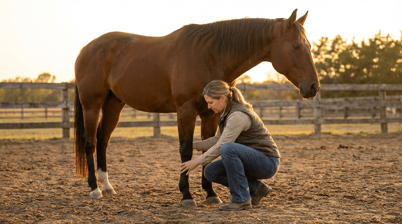
Palpation and Flexion
Systematic palpation of all four limbs — every joint, tendon, ligament, and hoof structure — is performed. The vet is assessing for heat, swelling, sensitivity to pressure, effusion (joint fluid), and asymmetry between limbs. Flexion tests — holding a limb in a flexed position for 60 to 90 seconds, then trotting the horse immediately away — apply concentrated stress to joints and soft tissues and can exacerbate subtle lameness. A positive flexion adds to the picture of where pain originates.
Dynamic Evaluation (In Hand and Under Saddle)
The horse is observed at the trot on a straight line, both directions, on hard ground. It is then worked on a lunge line on soft ground and on hard ground. The trot is the gait of choice for lameness evaluation because it is a symmetric two-beat gait — any asymmetry is easier to detect than in the three- or four-beat gaits. If the horse's primary use is under saddle and the lameness is not apparent in hand, the vet may request a ridden evaluation, which is especially valuable for back and sacroiliac pain.
Diagnostic Nerve Blocks (Perineural Analgesia)
Diagnostic nerve blocks are the cornerstone of equine lameness localization. A local anesthetic is injected around specific nerves or into specific joints to temporarily desensitize a region. After each block, the horse is re-evaluated at the trot. If the lameness improves by 70 percent or more after a particular block, that block has localized the pain to the region it desensitized.[3] Blocks are applied systematically from the foot upward — starting distally and working proximally — to progressively narrow the location of pain.
Common diagnostic blocks in a forelimb lameness workup:
- Palmar digital nerve block (PDN) — desensitizes the palmar third of the foot (navicular region)
- Abaxial sesamoid block — desensitizes the entire foot
- Low 4-point block (low palmar block) — desensitizes the fetlock and below
- High 4-point or High palmar block — desensitizes the pastern to the carpus
- Carpal block (intra-articular) — desensitizes the knee joint
Imaging: X-rays, Ultrasound, and MRI
Once lameness is localized to a region, diagnostic imaging provides structural information about the underlying pathology. The choice of imaging modality depends on what structures are being evaluated:
| Modality | Best For | Limitations | Approx. Cost (per region) |
|---|---|---|---|
| Radiography (X-rays) | Bone, joints, hoof capsule | Cannot image soft tissue; misses early bone changes | 150 to 400 dollars |
| Ultrasound | Tendons, ligaments, soft tissue, joint effusion | Cannot image inside hoof capsule; limited bone detail | 100 to 300 dollars |
| MRI (standing, low-field) | Entire foot, DDFT, navicular, coffin joint | Limited to distal limb; lower resolution than high-field | 800 to 1,500 dollars |
| MRI (high-field, general anesthesia) | Most detailed soft tissue imaging, any region | Requires anesthesia; significant cost | 2,000 to 4,000 dollars or more |
| Nuclear Scintigraphy (bone scan) | Active bone remodeling, stress fractures, SI pain | Limited soft tissue detail; radiation; referral facility | 1,500 to 3,000 dollars or more |
| CT scan | Complex fractures, distal limb detail, skull or spine | Requires anesthesia or specialized standing units; cost | 1,500 to 3,500 dollars or more |
Grading Lameness Severity
The American Association of Equine Practitioners (AAEP) lameness grading scale is the standard tool used by equine veterinarians to communicate the severity of lameness:[1]
| Grade | Description | Clinical Notes |
|---|---|---|
| 0 | No perceptible lameness under any circumstances | Normal horse |
| 1 | Difficult to observe; not consistently apparent | May only show under specific conditions (hard ground, circles, certain footing) |
| 2 | Difficult to observe at walk; consistently apparent at trot | Often the threshold at which diagnostic workup is initiated |
| 3 | Consistently observable at trot; occasionally at walk | Significant pain; usually warrants prompt treatment |
| 4 | Obvious lameness at walk | Severe pain; rest mandatory; often acute injury |
| 5 | Minimal weight bearing; unable to move, or completely non-weight bearing | Emergency — fracture, severe abscess, joint sepsis, or other acute crisis |
Treatment Options for Horse Lameness
Treatment of equine lameness depends entirely on the specific diagnosis. There is no universal lameness treatment, and applying the wrong therapy wastes money and time while the underlying problem progresses. Work with your equine vet to establish the diagnosis before committing to a treatment plan.
Farriery and Hoof Care
The relationship between lameness and hoof care cannot be overstated. The vast majority of foot pain problems are either caused by, worsened by, or treatable with appropriate farriery. For many lameness conditions, the farrier's contribution to treatment is equal to or greater than any drug or injection.
- Egg bar shoes: Extend the support surface behind the heel, reducing tension on the DDFT and navicular apparatus. A cornerstone of navicular syndrome management.
- Wedge pads and wedge shoes: Raise the heels to reduce DDFT tension; used in navicular syndrome and low-heel conformations.
- Wide web shoes and pads: Reduce sole pressure; useful in horses with thin soles, laminitis, and coffin bone remodeling.
- Heartbar shoes: Provide direct frog support to the coffin bone; used in laminitis management.
- Regular trimming intervals: Irregular or overly long trim intervals allow hoof imbalance to develop, directly contributing to lameness. Most horses need trimming every 6 to 8 weeks.
Joint Injections
Intra-articular injections are one of the most common treatments in equine lameness. They deliver anti-inflammatory agents directly into the affected joint, providing relief that is faster and more targeted than systemic medication. Common injection types include:
- Corticosteroids: The most potent anti-inflammatory option for joint injections. Highly effective for osteoarthritis management. Concerns about long-term cartilage effects at high doses exist but are largely mitigated with appropriate dosing and frequency (typically no more than 2 to 3 times per year per joint).
- Hyaluronic acid (HA): A naturally occurring joint fluid component that improves lubrication and has mild anti-inflammatory properties. Often combined with corticosteroids for synergistic effect.
- IRAP (Interleukin-1 Receptor Antagonist Protein): An autologous biological product made from the horse's own blood; specifically blocks interleukin-1, a key driver of cartilage degradation in OA.
- PRP (Platelet-Rich Plasma): Concentrated growth factors from the horse's own blood, used for both joint injections and soft tissue injuries. Evidence for efficacy is growing but variable.
- Stem cell therapy: Mesenchymal stem cells derived from bone marrow or fat tissue, used primarily for tendon and ligament injuries. Most evidence supports use in SDFT core lesions; less data for joint injections.
Systemic Medications
- NSAIDs (Phenylbutazone or Bute, Flunixin or Banamine, Firocoxib or Previcox): The primary systemic pain management option in horses. Phenylbutazone is the most widely used and cost-effective for musculoskeletal pain. All NSAIDs carry risk of gastric ulcers and right dorsal colitis with prolonged use; always use the lowest effective dose for the shortest necessary duration.
- Bisphosphonates (Tildren or tiludronate, Osphos or clodronate): IV or IM drugs that reduce bone resorption and pain in horses with navicular syndrome, coffin joint OA, and other bone-mediated pain conditions. Effect duration is typically 4 to 6 months.
- Gabapentin and other neuropathic pain agents: Used in some cases of chronic or neuropathic pain, particularly where standard therapies are insufficient.
Physical Therapy and Rehabilitation
Physical therapy and structured rehabilitation have become increasingly important components of equine lameness management, particularly for soft tissue injuries. Key modalities include:
- Controlled exercise programs: Many soft tissue injuries require a precisely graduated return to work. The horse must move enough to stimulate tendon fiber alignment and tissue remodeling, but not so much that it causes re-injury. Your vet will prescribe a specific exercise protocol.
- Therapeutic ultrasound: Applied to tendons and ligaments to promote healing and collagen alignment.
- Shockwave therapy (ESWT): High-energy acoustic waves delivered to injured tissue. Strong evidence for proximal suspensory desmitis and navicular syndrome.[5] Horses treated with shockwave should not be worked for at least 3 days post-treatment due to the pain-numbing effect.
- Cold therapy: Essential for acute injuries to reduce inflammation and swelling. Cold hosing, ice packs, and cold water boots all reduce tissue temperature and metabolic activity in injured areas.
- Controlled swimming and water treadmill: Facilities with aquatic treadmills allow horses to maintain cardiovascular fitness and promote limb movement without weight-bearing stress during recovery.
- Intralesional biological therapies: PRP and stem cells delivered directly into tendon or ligament lesions under ultrasound guidance to promote biological healing.
Surgical Options
Surgery is reserved for conditions that do not respond to medical management or where mechanical correction is required. Common surgical procedures in equine lameness include:
- Arthroscopy: Minimally invasive joint surgery for OCD fragment removal, cartilage debridement, and some fracture repairs. Recovery is generally faster than open joint surgery and outcomes are excellent for appropriate cases.
- Joint fusion (arthrodesis): Intentional surgical fusion of low-motion joints — particularly the distal tarsal joints (hock) and pastern — provides permanent pain relief by eliminating the painful joint surface. Horses with fused distal hock joints can return to full work and often become completely sound.
- Neurectomy (palmar digital neurectomy): Surgical severing of the palmar digital nerves to desensitize the back of the foot. Used as a management tool for navicular syndrome when other treatments are insufficient. Requires ongoing careful hoof care and carries risks that must be discussed with your veterinarian.[2]
Costs of Equine Lameness Workup and Treatment
Horse lameness is notoriously expensive to diagnose and treat. Understanding what costs to expect helps with planning and ensures you can have an informed conversation with your veterinarian about diagnostic and treatment priorities.
Diagnostic Costs
| Service | Typical Cost Range | Notes |
|---|---|---|
| Farm call or farm visit fee | 75 to 200 dollars | Plus mileage in rural areas; higher in high-cost states |
| Basic lameness examination | 100 to 250 dollars | Palpation, flexion tests, movement evaluation |
| Diagnostic nerve blocks (per block) | 50 to 150 dollars | Typical workup uses 2 to 6 blocks |
| Radiographs (per view) | 30 to 60 dollars | A typical foot series = 4 views; 120 to 240 dollars |
| Ultrasound examination (per region) | 100 to 300 dollars | Digital and limb ultrasound on site |
| Standing MRI (referral) | 800 to 1,800 dollars | Low-field, distal limb; no anesthesia required |
| High-field MRI (anesthesia) | 2,500 to 4,500 dollars | Best detail; any region; referral center only |
| Nuclear scintigraphy (bone scan) | 1,500 to 3,000 dollars | Referral facility; excellent for diffuse or obscure pain |
| Complete lameness workup (typical) | 500 to 1,500 dollars | Blocks plus radiographs plus ultrasound on one limb |
Treatment Costs
| Treatment | Typical Cost Range | Frequency or Notes |
|---|---|---|
| Single joint injection (corticosteroid plus HA) | 150 to 400 dollars | Every 3 to 12 months depending on joint and response |
| Bisphosphonate treatment (Tildren or Osphos) | 400 to 800 dollars | Every 4 to 6 months for navicular syndrome |
| IRAP series (3 to 5 injections) | 600 to 1,500 dollars | Blood draw plus processing plus injection fees |
| PRP preparation and injection | 300 to 700 dollars | Per treatment; may need 2 to 3 treatments |
| Stem cell therapy | 1,500 to 3,500 dollars | Bone marrow harvest plus culture plus injection |
| Therapeutic shoeing (specialty shoes) | 100 to 400 dollars per shoeing | Every 6 to 8 weeks ongoing |
| Shockwave therapy (3-treatment course) | 300 to 900 dollars | 3 sessions 3 weeks apart is typical protocol |
| OCD arthroscopy | 2,500 to 5,000 dollars or more | Includes anesthesia, surgery, hospitalization |
| Distal tarsal (hock) arthrodesis | 2,000 to 5,000 dollars | Chemical or surgical; very effective for bone spavin |
| Neurectomy | 1,500 to 3,000 dollars | Palmar digital; effective for navicular when indicated |
Lameness in Different Disciplines and Breeds
Lameness patterns vary significantly by discipline and breed, reflecting the biomechanical demands placed on horses in different activities.
Racehorses (Thoroughbred and Quarter Horse)
Racing places extraordinary demands on the musculoskeletal system at high speed and under significant fatigue. Common lameness issues include: soft tissue injuries of the lower limb (SDFT bowing is classic), condylar and sesamoid fractures (catastrophic breakdown injuries), bucked shins (dorsal cortical metacarpal stress fractures) in young horses entering training, and fetlock and pastern joint OA from repetitive high-speed concussion. Racing regulatory bodies in each state have specific veterinary oversight requirements, and horses must pass a soundness evaluation before racing.
Warmbloods and Sport Horses
Dressage, show jumping, eventing, and combined driving horses are heavily represented among horses with proximal suspensory desmitis, hock osteoarthritis (bone spavin), and back and sacroiliac pain. The collected movements of dressage place intense demands on the hindlimbs and back. Jumping concentrates enormous forces on the forelimbs at landing. Navicular syndrome is common in all disciplines. OCD is frequently diagnosed in young Warmbloods intended for sport horse careers and can affect saleability and career length significantly.
Quarter Horses and Western Performance Horses
Cutting, reining, barrel racing, and roping place unique stresses on the musculoskeletal system. Deep digital flexor and navicular pathology are common in reining horses due to the extreme sliding stops and tight spins. Stifle problems — particularly OCD and soft tissue injuries — are more common in cutting horses. Barrel horses have a higher incidence of hindlimb lameness from the tight turns demanded by the pattern.
Draft Horses
Draft breeds have a unique lameness profile driven by their massive size and the work they are asked to do. Scratches (pastern dermatitis) and chronic progressive lymphedema (a heritable lymphatic disorder in some European draft breeds) are unique to these horses. Joint disease is common simply due to the enormous weight bearing on each limb. Stringhalt — a neurological condition causing exaggerated hindlimb flexion at the walk — is more frequently reported in Australian draft horses but occurs across breeds.
Ponies and Miniature Horses
Ponies and miniature horses have disproportionately high rates of laminitis due to their tendency toward obesity and insulin dysregulation. Easy-keeper breeds require careful dietary management — particularly in spring when pasture NSC levels are high — to prevent recurrent laminitis episodes.
Seasonal Considerations in Equine Lameness
Lameness patterns shift predictably with the seasons, and awareness of these patterns helps horse owners anticipate and prepare.
Spring
Spring is the highest-risk season for pasture-associated and endocrine laminitis. As grass growth surges and NSC content peaks in rapidly growing cool-season grasses, EMS and PPID horses are acutely at risk. Monitor at-risk horses closely in April and May. Restrict grazing time, use grazing muzzles, and avoid turnout during periods of rapid grass growth — NSC concentrations peak in the evening and remain high through morning. Increase routine farrier visits to monthly for horses with a history of laminitis.
Summer
Hard, dry summer ground increases concussive forces and exacerbates navicular disease, foot soreness, and splints in horses worked frequently on hard surfaces. Horses sweating heavily in summer heat lose significant electrolytes, which can affect muscle function. Dry hooves are more brittle and prone to cracking; regular farrier care and hoof conditioner use are important.
Fall
Fall is the second peak season for laminitis in EMS horses as cool nights and warm days drive a second flush of grass NSC accumulation. Horses being brought back into training after summer pasture turnout may develop exercise-associated lameness from soft tissue stress on decompensated structures. Shoeing changes from summer barefoot to shod status can unmask navicular syndrome.
Winter
Cold temperatures cause horses to move less, leading to stiffness and loss of conditioning that can worsen chronic lameness conditions. Horses with arthritis are often more stiff and sore in cold weather; increased NSAID use and supplemental heat (blankets, warm housing) may be needed. Ice and frozen ground create traction hazards that can result in slips and acute injuries. Horses in deep mud during muddy winter periods are at risk for dermatitis, white line disease, and secondary hoof problems that predispose to lameness.
When to Call Your Equine Veterinarian
Some lameness presentations require immediate veterinary attention. Knowing which signs warrant an emergency call versus a scheduled appointment could save your horse's life or career.
Emergency — Call Immediately
- Non-weight-bearing (Grade 5) lameness on any limb
- Obvious swelling, heat, or deformity suggesting fracture or joint injury
- Any penetrating wound near a joint or tendon sheath — joint infection (septic arthritis) is a surgical emergency
- Acute, severe laminitis — reluctance to move, hot hooves, shifting weight, digital pulse bounding
- Signs of heel bulb or coronary band penetration or rupture
- Horse down and unable to rise due to limb pain
- Suspected fracture — any abrupt onset severe lameness after a sudden movement, fall, or collision
Schedule Within 24 to 48 Hours
- Moderate (Grade 3 to 4) lameness that does not resolve within a day of rest
- Suspected hoof abscess that has not drained within 24 to 48 hours of home soaking and poultice
- New onset lameness in a competition horse with an event upcoming
- Any lameness in a horse in the third trimester of pregnancy
- Lameness accompanied by significant heat, swelling, or joint effusion
Schedule for a Routine Appointment
- Subtle, consistent Grade 1 to 2 lameness without obvious cause
- Performance decline, reluctance to collect or engage the hindquarters, or subtle asymmetry noticed under saddle
- Recurring lameness in the same limb that resolves with rest but returns with work
- Annual soundness examination or pre-purchase examination
- Follow-up imaging to assess healing of known soft tissue injury
Frequently Asked Questions About Horse Lameness
How long does it take for a lame horse to recover?
Recovery time varies enormously depending on the cause. A simple hoof abscess may resolve in 3 to 7 days. A joint injection for mild arthritis may provide relief within 1 to 2 weeks with return to full work. A soft tissue injury like a suspensory ligament lesion may require 6 to 12 months of rest and rehabilitation before return to competition. Severe laminitis may require 12 to 24 months of intensive management, and some horses never return to their previous level of work. Your veterinarian will give you a prognosis and expected timeline based on the specific diagnosis and severity of disease.
Should I give my horse bute for lameness without calling the vet?
Short-term use of phenylbutazone (bute) to manage acute, mild lameness while awaiting a veterinary appointment is reasonable for horses with a known history of musculoskeletal issues. However, masking pain with NSAIDs without identifying the underlying cause risks allowing an acute injury to progress while the horse appears to feel better. Bute given before a lameness evaluation also temporarily suppresses pain signals, making nerve block interpretation more difficult. Never give bute to a horse you suspect has a fracture, a joint infection, or colic without veterinary guidance.
What is a pre-purchase examination (PPE)?
A pre-purchase examination is a veterinary examination performed before buying a horse to evaluate its health, soundness, and suitability for the intended use. A comprehensive PPE includes a full lameness examination with nerve blocks, radiographs of key joints, blood work, and often an upper airway scope. The vet reports findings objectively and provides an opinion on whether identified conditions are likely to affect the horse's ability to perform the buyer's intended job. A PPE is not a guarantee of future soundness but is the single best investment you can make before purchasing any horse intended for performance or significant value.
Can a lame horse still be ridden?
This depends entirely on the cause and grade of lameness. A horse with Grade 1 lameness from mild arthritis managed with joint injections may be appropriate for continued work — in fact, controlled exercise is often therapeutic for arthritic joints. A horse with Grade 3 or higher lameness should not be ridden until the cause is identified and appropriately treated. Riding a significantly lame horse risks worsening the underlying injury, causing compensatory damage in other limbs, and compromising the horse's welfare. When in doubt, rest and consult your veterinarian before riding.
What is bone spavin?
Bone spavin is the common term for distal tarsal osteoarthritis — degenerative joint disease of the small, low-motion joints at the bottom of the hock. It is one of the most common causes of hindlimb lameness in working horses and is particularly prevalent in Warmbloods, draft breeds, and horses that do a lot of collected work. Bone spavin causes pain during the active phase of cartilage erosion; once the joint surfaces have fully eroded and the joint fuses naturally, many horses become spontaneously sound. Treatment options include joint injections, exercise modification, and surgical or chemical arthrodesis to accelerate fusion and resolution of pain.
How can I prevent lameness in my horse?
No approach guarantees a sound horse, but the following practices significantly reduce lameness risk: maintain a regular farrier schedule (every 6 to 8 weeks), ensure a balanced diet with appropriate trace minerals, introduce new training demands gradually without rapid workload increases, warm up and cool down properly before and after exercise, maintain footing quality in the areas where your horse works, keep the horse at a healthy body weight, schedule annual veterinary wellness exams to catch early structural changes, and address lameness signs early before minor issues become major ones.
What does it mean if my horse is sore in both front feet equally?
Bilateral forelimb soreness — where both feet are equally affected — can be challenging to detect as a lameness because the horse may not nod dramatically on either limb. Instead, these horses typically have a shortened, choppy, or pottery gait, may stumble frequently, and may resist working on circles. Bilateral navicular syndrome, bilateral laminitis, and bilateral coffin joint arthritis are common diagnoses. These horses require a full lameness workup including diagnostic blocks and imaging of both feet, and treatment is typically directed at both limbs simultaneously.
Summary: Managing Your Horse's Soundness
Horse lameness is a complex, multifactorial challenge that affects horses in every discipline, at every level of competition, and in every region of the country. The key principles of successful lameness management are:
- Know your horse's normal: Daily observation helps you catch subtle changes before they become serious injuries.
- Act early: Early lameness intervention consistently produces better outcomes than delayed treatment. Do not wait for walk lameness before calling your vet.
- Get an accurate diagnosis: Treatment without diagnosis is guesswork. A systematic lameness workup including nerve blocks and imaging is the foundation of appropriate treatment selection.
- Follow the rehabilitation protocol: Especially for soft tissue injuries, the return-to-work protocol prescribed by your vet is not a suggestion — it is the difference between a horse that comes back sound and one that re-injures.
- Maintain the partnership with your farrier and vet: Regular farriery and annual veterinary exams are preventive investments, not optional expenses.
- Respect your horse's signals: Horses that resist certain movements, show behavioral changes, or underperform are often expressing pain. Investigate before attributing changes to attitude or training issues.
Find an Equine Veterinarian Near You
Horse lameness diagnosis and treatment requires an experienced equine veterinarian with appropriate diagnostic equipment — including portable digital radiography, ultrasound, and ideally access to advanced imaging like MRI and bone scan at a referral center. Not all rural large animal practices have the same level of equine expertise, and finding the right vet for your horse and discipline matters.
FarmVetGuide is the most comprehensive directory of large animal and equine veterinary practices in the United States, with over 9,500 listings across all 50 states. You can filter by species treated (equine), emergency availability, mobile service, and geographic location to find board-certified equine practitioners, ambulatory vets who come to your farm, and full-service equine hospitals near you.
Whether your horse needs a routine lameness evaluation, an emergency farm call, or a referral to an equine surgical center, start your search at FarmVetGuide.com — the trusted resource for horse owners and farm managers across America.
Sources & References
This guide references peer-reviewed research and guidelines from leading veterinary organizations. All medical information has been reviewed for accuracy against these authoritative sources.
- American Association of Equine Practitioners (AAEP) — Lameness Exams: Evaluating the Lame Horse. Last accessed March 2026.
- Merck Veterinary Manual — Navicular Syndrome in Horses: Pathophysiology, Diagnosis, and Management. Last accessed March 2026.
- Merck Veterinary Manual — Diagnostic Anesthesia for Lameness Localization in Horses: Perineural and Intra-articular Blocks. Last accessed March 2026.
- Merck Veterinary Manual — Laminitis in Horses: Causes, Obel Grading, Coffin Bone Rotation, and Treatment. Last accessed March 2026.
- Merck Veterinary Manual — The Lameness Examination in Horses: Flexion Tests, Nerve Blocks, and Imaging. Last accessed March 2026.
- American Association of Equine Practitioners (AAEP) — Equine Lameness: Clinical Judgment Meets Advanced Diagnostic Imaging. Last accessed March 2026.
- PMC / PeerJ — A Modified Obel Method for the Severity Scoring of Endocrinopathic Equine Laminitis. Last accessed March 2026.